


Dermatological Treatments
Nevus (Mole) Monitoring and Removal
Being able to read the story of a mole is the most powerful tool of early diagnosis.
Overview
A nevus, commonly known as a mole, is a benign skin lesion made up of pigment cells. Most moles remain harmless for life; however, some change in colour, border, size or surface and require monitoring.
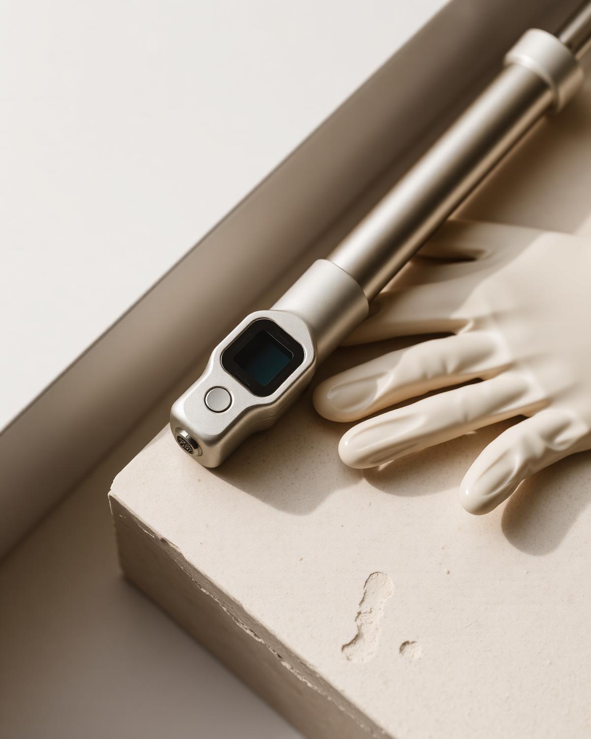
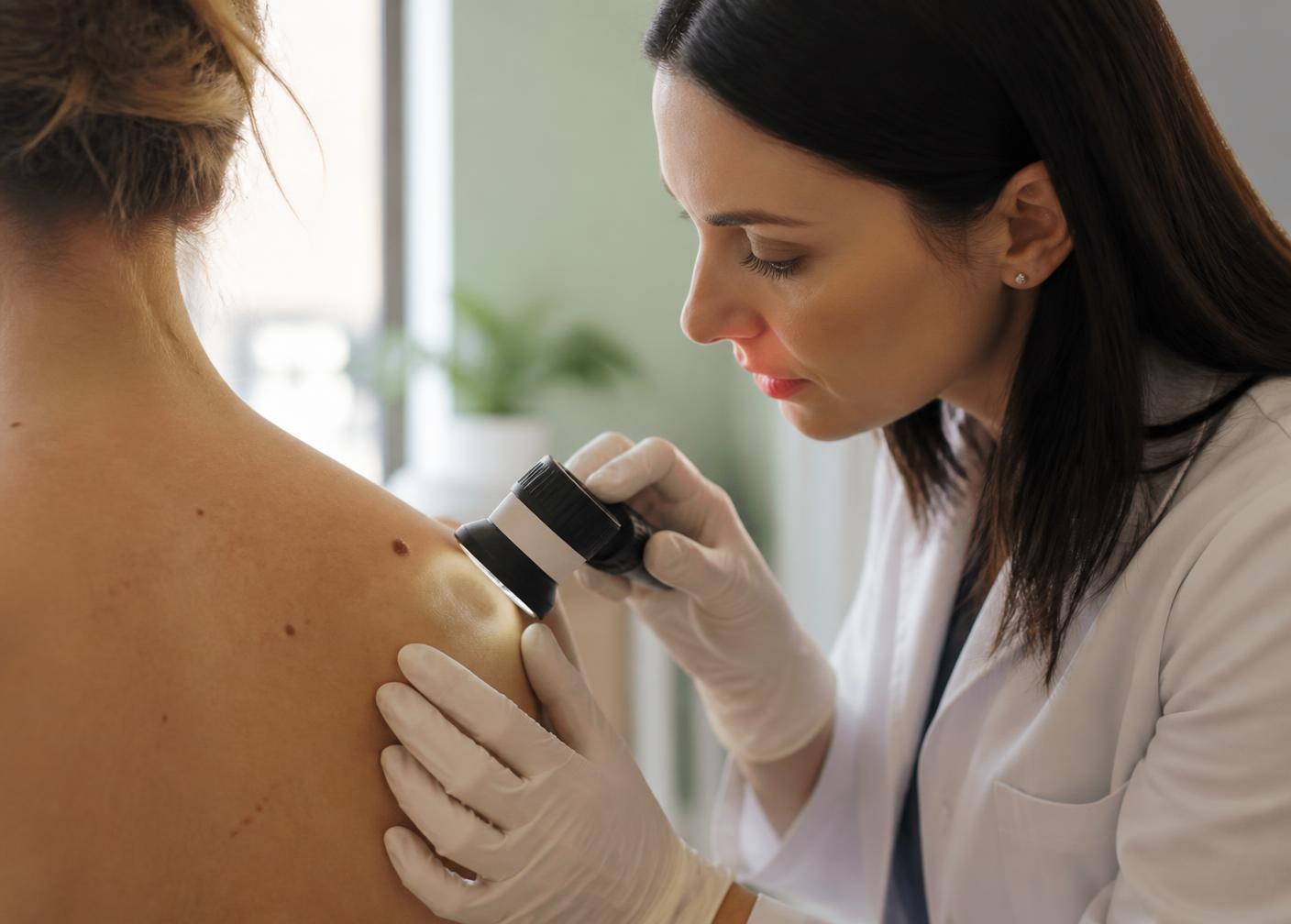
The assessment is carried out with a dermatoscope. This device magnifies the pigment network beneath the skin surface, revealing structural clues invisible to the naked eye. In people with many moles, digital mapping allows the same lesion to be compared over time.
Moles that look suspicious or show irritation, bleeding or rapid growth are removed surgically under local anaesthesia and sent for pathological examination. For moles removed for cosmetic reasons, the method is chosen with scar control in mind.
Who is it for?
- People with many moles who want regular screening
- Those with a family history of skin cancer
- Those who notice a change in a mole's colour, border or size
- Those with moles constantly irritated by clothing, shaving or jewellery
- Fair-skinned people prone to freckling and sunburn
Treatment Information
- Examination time
- 20 – 30 minutes
- Removal time
- 15 – 30 minutes
- Follow-up interval
- 6 – 12 months
- Pathology result
- 7 – 10 days

Approach
“Being able to read the story of a mole is the most powerful tool of early diagnosis.”
Dr. Sera Kayhan · Dermatologist
Process
How does the treatment proceed?
- 01
Dermoscopy
A detailed full-body mole examination is performed with a dermatoscope.
- 02
Mapping
Suspicious lesions are photographed and recorded digitally.
- 03
Decision
Whether monitoring or surgical removal is needed is decided together, based on the findings.
- 04
Removal and pathology
The relevant lesion is removed under local anaesthesia and examined pathologically.
FAQ
About Nevus (Mole) Monitoring and Removal
Is having a mole removed harmful?+
No. Mole removal performed with the right indication, proper technique and pathological examination is safe; on the contrary, it enables early diagnosis in suspicious lesions.
Which moles should be checked?+
Moles that are asymmetric, have irregular borders, contain more than one colour, are larger than 6 mm or change over time should be assessed as a priority.
Will there be a scar after the procedure?+
A fine scar may form after surgical removal; depending on the area and suture technique, it fades noticeably over time.
How often should I have a mole check?+
Once a year for those without risk factors; every 6 months for people with many moles or a family history of melanoma.
Can moles be removed with a laser?+
Laser is not preferred for suspicious naevi, because no tissue sample can be obtained for pathological examination. The decision is always based on dermoscopic findings.
Dermatological Treatments
Acne and Scar Treatment
A plan that stops active acne and gradually repairs scars.
View →Rosacea
A control plan for persistent redness, visible capillaries and sensitivity.
View →Diagnosis and Treatment of Skin Diseases
Skin health, barrier repair, and revitalization protocols.
View →Pigmentation Treatment
Personalized protocol for melasma, sun spots, and post-inflammatory marks.
View →Nail Disorders and Nail Removal
Diagnosis and treatment for fungal infection, ingrown nails and colour changes.
View →